Swine producers and veterinarians need both nucleic acid- and antibody-based tests to monitor PRRSV. PRRSV RT-PCR reflects virus circulation at the time of sample collection, whereas antibodies reveal the history of infection and immunity.
January 2, 2018
By Marisa Rotolo, Luis Giménez-Lirola, Ronaldo Magtoto, Ju Ji, Chong Wang, David Baum, Rodger Main and Jeff Zimmerman, Iowa State University
Porcine reproductive and respiratory syndrome virus infection (or vaccination) stimulates the pig’s immune system to produce three types of antibodies: IgM appears within seven days of infection, IgA after seven days, and IgG at nine to 10 days of infection. The current PRRSV oral fluid ELISA is designed to detect IgG because it is present at a higher concentration and for a longer period of time than IgM or IgA. The PRRSV oral fluid IgG ELISA is very effective at monitoring maternal antibody levels in young pigs and the PRRSV immune status of growing pig populations.
The new PRRSV oral fluid IgM-IgA ELISA is designed to simultaneously detect both IgM and IgA, but not IgG. The two advantages of this new assay is that (1) it is able to detect active infection in piglets with circulating maternal IgG antibody and (2) can serve as a confirmatory assay in the event of “unexpected” results from other PRRSV tests, for example PRRSV RT-PCR or IgG ELISA.
Detection of PRRSV infection in pigs with colostral IgG
Sows that have been infected or vaccinated with PRRSV provide IgG to their piglets in their colostrum. Depending on the amount of colostral PRRSV IgG provided by the sow, piglet serum and oral fluid samples will test positive on the PRRSV IgG ELISA for four to eight weeks post-weaning. Thus, in weaned pigs, a PRRSV IgG ELISA positive test result could be due either to maternal IgG or IgG produced in response to PRRSV infection. How do you know which is which?
Here is the answer: Colostral IgM and IgA do not enter the piglets’ circulation and/or oral fluids. Therefore, IgM and/or IgA in oral fluids are only present if the pig(s) have been infected with PRRSV. We investigated this idea in two studies.
Study 1 (Experimental study): In Study 1, 12 PRRSV-naïve pigs (100 pounds each) were housed in separate pens under experimental conditions. Each pig was vaccinated with a PRRS modified-live vaccine on Day Post-Vaccination 0. Oral fluids were collected from each pig from days post vaccination -7 to 42. In total, 600 oral fluid samples were collected, randomized and tested with the PRRSV IgG ELISA and the PRRSV IgM-IgA ELISA. Test results are shown in Figure 1. Note that the PRRSV IgM-IgA ELISA yielded earlier detection than the IgG ELISA, then gradually declined over time. The results confirmed that IgM-IgA were rapidly present at detectable levels in oral fluids following PRRSV exposure.
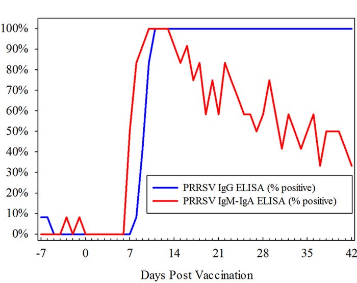
Figure 1: Individual pig oral fluid antibody test results from 12 animals vaccinated with MLV PRRSV vaccine under experimental conditions.
Study 2 (Field study): In Study 2, pen-based oral fluids were collected on three commercial wean-to-finish sites (A, B, C). Each site had three barns (1,000 pigs per barn, 36 pens per barn). Every pen in every barn was sampled every week for eight weeks beginning one week post-placement. In total, 2,899 pen-based oral fluid samples were collected, randomized and then tested by PRRSV RT-PCR, the PRRSV oral fluid IgG ELISA, and the PRRSV oral fluid IgM-IgA ELISA.
Sites A and B were PRRSV RT-PCR and PRRSV IgM-IgA ELISA negative throughout the sampling period. Approximately 85% of oral fluid samples from these sites were PRRSV IgG ELISA positive at the initial sampling, with declining rates of positivity, thereafter.
On Site C, ~5% of samples were PRRSV RT-PCR positive at the initial sampling. Among these samples, 100% were PRRSV IgG ELISA positive and 0% were PRRSV IgM-IgA ELISA positive. This scenario suggested an active PRRSV infection in the sow herd (high levels of colostral antibody) and the recent introduction of PRRSV into the weaned pig population (low level of PRRSV RT-PCR samples). As shown in Figure 2, the rate of PRRSV RT-PCR positivity increased steadily over time, followed shortly thereafter by an increase in PRRSV IgM-IgA ELISA positivity. The PRRSV IgG ELISA results showed a decline in the percent of positive samples until four weeks post-placement, followed by an increase that mirrored the IgM-IgA ELISA response.
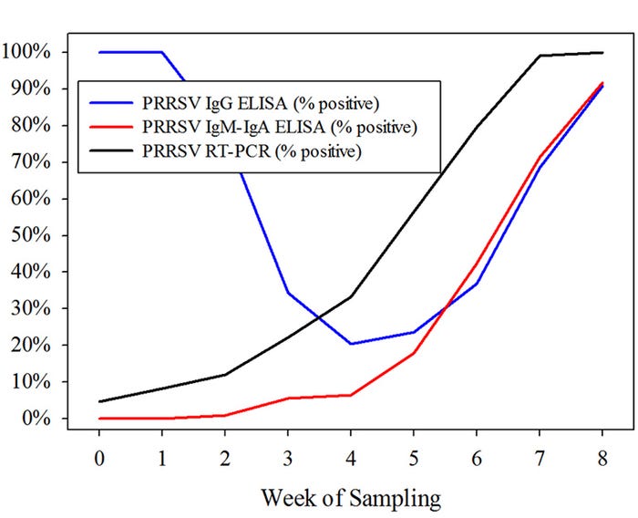
Figure 2: Pen-based oral fluid test results from three wean-to-finish barns (36 pens per barn). Despite the presence of PRRSV IgG colostral antibody, PRRSV spreads through the site, as indicated by PRRSV RT-PCR and IgM-IgA ELISA results.
Implications/conclusions
Swine producers and veterinarians need both nucleic acid- and antibody-based tests to monitor PRRSV. PRRSV RT-PCR reflects virus circulation at the time of sample collection, whereas antibodies reveal the history of infection and immunity. The current PRRSV IgG-based ELISAs cannot differentiate maternal antibody from the pig’s response to infection, but the new IgM-IgA oral fluid ELISA could detect PRRSV infection in weaned pigs — even pigs with maternal antibody. The purpose of the IgM-IgA oral fluid ELISA is to provide producers and veterinarians the ability to differentiate antibody responses in pigs. This assay may also be used as a confirmatory assay. Timing and duration of detectable antibody are important factors to consider when using or interpreting PRRSV IgM-IgA Elisa antibody results (Figure 1).
Future research is needed to determine if the PRRSV IgM-IgA oral fluid ELISA may have a targeted pen or lot-level vaccination compliance application in pigs coming from seropositive sows. The PRRSV IgM-IgA oral fluid ELISA is currently available at the Iowa State University Veterinary Diagnostic Laboratory.
You May Also Like
