
Anaplasmosis
Anaplasmosis is a tick-borne rickettsial disease caused by Anaplasma marginale. The disease is endemic in many parts of the world including the US. Prevalence is higher in the southern US and is thought to be lower in the northern US and Canada; however, anaplasmosis is still being reported for all of these areas often in proximity to river areas especially as cattle begin to travel.
Transmission is typically through ticks of Dermacentor spp. and mechanical through transmission by needles and/or surgical instruments. Biting insects are also involved in transmission.
Cattle of all ages can be infected with anaplasmosis with severity of clinical disease related to the age of the animal. Mortality is most common in animals greater than two years old. Clinical disease is often reported late August through frost.
| Age | Clinical Signs |
|---|---|
| <6 months old | Rare, but can become infected |
| 6 – 12 months old | Mild subclinical disease |
| 1 – 2 years old | Acute but rarely fatal disease |
| 2 years old | Acute and often fatal disease |
| >2 years old | Present initially with anorexia, off feed, constipated with pale mucous membranes. Dyspnea and exercise intolerance may become apparent, with marked aggression common. More often present as “sudden death”. After 3 years of age 30-50% of clinical anaplasmosis cattle die if untreated. |
Lesions Associated with Anaplasmosis
- Pale mucus membranes
- Marked icterus [yellow to golden hues of mucus membranes, conjunctiva, and areas of unhaired skin]
- Thin, watery blood
- PCV can and usually will be extremely low
- Enlarged, pulpy to mushy spleen, with prominent follicles
- Hepatomegaly with yellow-orange discoloration and rounding of edges
Stages of Anaplasmosis
- Incubation stage – begins at infection. Average incubation stages are 3-8 weeks, but wide ranges have been seen. Animals appear clinically normal at this stage.
- Developmental stage – lasts 4-9 days, characteristic clinical signs occur.
- Forced movement and excitement can result in death due to anoxia
- Antibiotic treatments do little to affect the outcome of disease during late developmental and early convalescent stages. Death typically occurs during the late developmental or early convalescent stages.
- Convalescent Stage – cattle that survive clinical disease lose weight, abort calves and recover slowly over a 2-3 month period. Increased erythropoiesis is typically observed in this time frame.
- Carrier Stage – Unless medicated appropriately cattle that recover from anaplasmosis remain carriers for life. During the carrier stage animals do not exhibit clinical signs.
Diagnosis
Whole blood collected in EDTA or heparin tubes from clinically affected animals is the diagnostic sample of choice. However, the ISU VDL has an Anaplasma spp. PCR available with valid sample types: EDTA blood, heparinized blood, or spleen. Additionally, a concurrent submission of air-dried, unstained, blood smear preparations is also encouraged. Serology can be used to evaluate for possible infection, or used on a herd basis to evaluate prevalence.
Collection of formalin-fixed and fresh tissue for differential diagnosis is preferred in most instances, particularly in areas where endemic infection is uncommon.
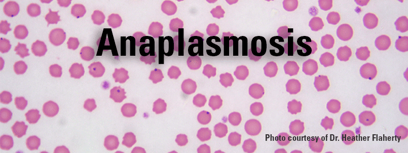
In the clinical setting diagnosis on a stained blood smear using Diff-Quik, Giemsa, or Wright’s stains for organisms, will provide a rapid diagnosis.
Prevention
Vaccination does work to prevent acute expression of disease; however, it does not prevent infection or the development of carrier animals. Nutrition and environmental stress are important factors in suspect herds. Additionally, reducing vector transmission (biting insects) and mechanical transmission of instruments used to work cattle are important.