A Giant Step Forward
The previous microscope researchers in the Department of Biomedical Science and others at Iowa State University used was pretty amazing.
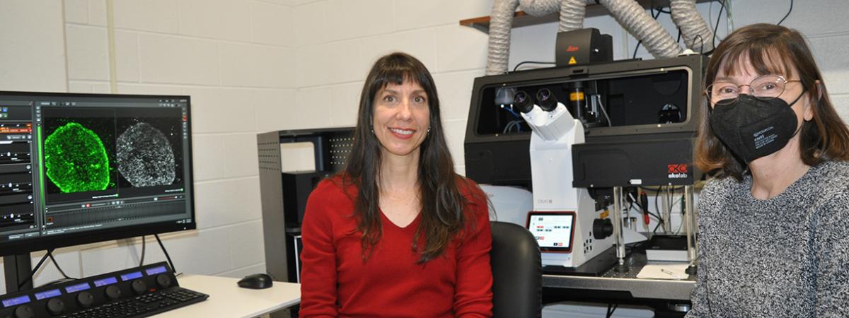
But the new stimulated emission depletion (STED) super-resolution microscopy system is in a class all its own.
“Advances in technology often provide substantial improvements in research capabilities, but seldom is there an advancement that literally opens new vistas of research for a wide range of disciplines, including human, animal, plant and microbial research alike,” said Dr. Michael Kimber, professor of chair of the Department of Biomedical Sciences.
The new STED microscopy system was funded through a grant from the Roy J. Carver Charitable Trust of Muscatine, Iowa, and additional support from Iowa State’s Office of Biotechnology. The instrumentation was installed recently in the College of Veterinary Medicine and is administered by the Office of Biotechnology. It can be utilized by researchers and students in biomedical sciences, biochemistry, biophysics, molecular biology, genetics, development and cell biology, as well as other disciplines across campus.
Previously campus researchers were limited by the physics of light diffraction through glass lenses. The resolution would be curtailed at around 200 nanometers, making it impossible for traditional fluorescent microscopy methods to discern subcellular structures and organelles.
That won’t be the case with the new STED microscopy system. In STED microscopy, a donut-shaped depletion beam is applied to a confocal laser scanning microscopy signal to produce the sharper resolution.
“The STED system will allow researchers to look at 30 to 50 nanometers,” said Margie Carter, manager of the STED system with the Office of Biotechnology. “Now researchers will be able to see much more than they ever could before.”
Jeanne Serb, director of the Office of Biotechnology, says there’s another advantage to the STED system.
“Not only will researchers be able to go view cellular structure at a much finer scale, but the STED system will allow them to keep cell lines alive when they are looking at them through the microscope,” she said. “There was a gap before where we couldn’t look inside living cells.”
This will aid Iowa State researchers in vaccine development. Other research projects that could potentially benefit from the STED system including research to treat parasitic diseases such as malaria, the discovery of new therapeutic targets for inflammatory diseases, testing how to overcome drug-resistant pathogens such as tuberculosis and COVID-19, better understanding birth defects, neurological disorders, male infertility in humans and animals and the aging process’s impact on the heart and liver, and plant metabolism to improve human food, animal
feed, bioenergy and bioproducts. “This is a game changer for our researchers,” Serb said. “Before they had to make assumptions because they really couldn’t see what was going on. Now they will be able to make observations in real time.”
Serb says the STED system will also change the type of research can be conducted at Iowa State.
“Researchers will be able to ask, and potentially answer, more probing questions than they could ever before,” she said.
April 2022
